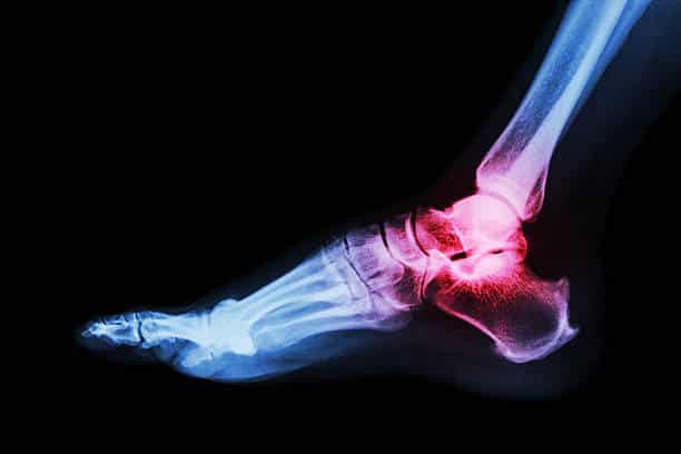
Sinus Tarsi Syndrome
The purpose of this case study is to assist our graduates to fully reflect on a client that they have seen throughout the week. This will also form part of information that we can distribute to clients where they can read up on real life cases where we have been able to help clients and allow them to be pain free! Clients are refered to as Mr.X/Ms.X to keep their privacy.
TITLE OF BLOG: Sinus Tarsi Syndrome
Section 1: About your client and how you diagnosed the condition (think about how they presented, what subjective and objective information did you gather to help you diagnose?)
Client presented with lateral ankle discomfort, located anterior and slightly inferior to the lateral malleolus (ball on the outside of the ankle). This client mentioned they have sprained their ankle a number of times and did not seek rehabilitation, However, this time has lingered so decided to seek treatment.
Recurrent ankle sprains are quite common in the cause of STS. STS is usually experienced post a traumatic incident or with reduced stability around the ankle – such as the result following a number of previous ankle sprains. One of many reasons to seek appropriate rehabilitation following an ankle sprain.
On observation, the client had quite pronated, or flat feet which can increase the risk of sinus tarsi syndrome as this closes down the space causing a compressive, irritating effect.
Ankle eversion (turning ankle/ foot outwards) also provoked symptoms, whilst inversion (turning ankle inwards) was an easing movement and reduced symptoms.
Section 2: Your diagnosis and about the condition (what is your possible diagnosis?)
Possible diagnosis: Sinus Tarsi syndrome
Pathophysiology background:
This pathology is mostly a result of synovitis and infiltration of fibrotic tissue into the sinus tarsi space due to an instability of the subtalar joint, caused by ligamentous injuries.
The sinus tarsi syndrome can also occur as a compression injury, for example to people who have flat or pronated feet. The talus and calcaneus are pressed together as a result of the deformation. This causes bone to bone contact of the talus and calcaneus, with inflammation or arthritis in the sinus
Section 3: Differential Diagnosis (what is another condition to consider and why?)
Chronic ankle instability, peroneal tenosynovitis, ankle sprain, calcaneal/ talus fracture.
Section 4: Treatment (what did you do and why?)
Initial management consisted of establishing aggravating patterns. For this individual, uneven surfaces and standing for long periods were these.
Education was provided to reduce aggravating patterns. I also taped the client to allow a greater arch in the foot, as well as recommending a generic arched shoe sole to reduce the flat foot and therefore the aggravating pattern of the condition.
This was paired with some initial strengthening of surrounding tissue such as the arch of the foot to reinforce a more appropriate foot position.
Section 5: Plan (where to go from here? How many sessions might they need? What’s the goal?)
The goal for this individual was to be skiing in 6 weeks time. The first two weeks were spent continuing to reduce the aggravating factors and strengthening surrounding tissue, including the foot intrinsic muscles to help support a better arch. We then progressively strengthened further around the ankle, including the calf and surrounding stabilising muscles. This followed into working into ankle eversion (being an initially aggravating movement), as this is an important movement when skiing. Plenty of balance and proprioception exercises were also completed to restore appropriate movement patterns at the ankle.
Final stages involved plyometric movements into ankle eversion to ensure they were ready for the slopes!

