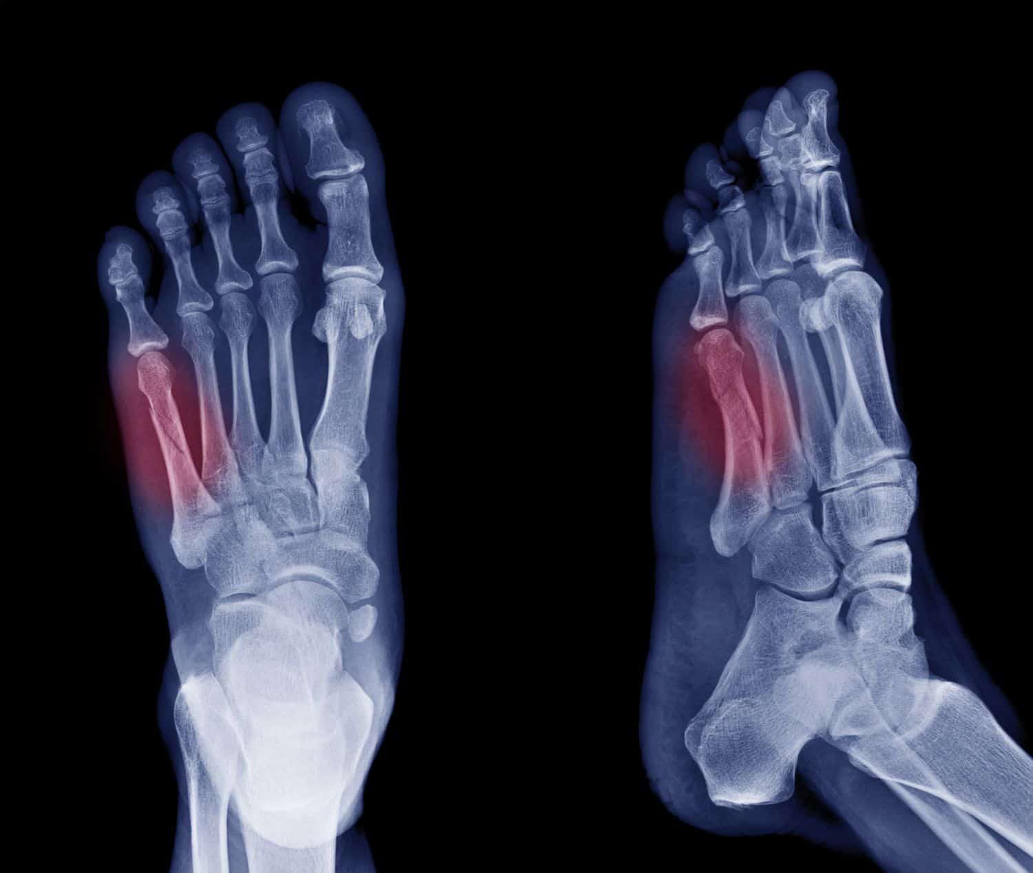
Metatarsal stress fracture
CASE STUDY REFLECTION BLOG POST: Metatarsal stress fracture
Section 1: About your client and how you diagnosed the condition (think about how they presented, what subjective and objective information did you gather to help you diagnose?)
Mrs. X presented to us in a CAM boot and crutches following a confirmed fracture in her 3rd metatarsal. The fracture occurred 5 weeks ago, and Mrs. X has been on crutches for the past week. She was unsure of the mechanism of injury however believes she sustained the fracture while playing football. She cannot remember an incident that may have caused her fracture, however has noted that she had been playing 2 matches in the 1 day a few weeks prior to the onset of pain. She recalls pain on weightbearing as well as swelling in the midfoot region in the acute phase post injury, however Mrs. X now has 0/10 pain both at rest and on palpation. Mrs. X has not been consistently wearing her CAM boot over the course of her injury, however has been wearing it consistently over the last week. She is required to ambulate on crutches for at least 1 more week as per her doctors orders to allow for fracture healing.
Upon examination, Mrs. X had no pain upon palpation over the 3rd metatarsal as mentioned above. She had reduced strength and ROM on her effected side and reported feeling ‘weaker and stiffer’ in her ankle. Due to her current protected weightbearing status we did not conduct any other objective assessments.
Section 2: Your diagnosis and about the condition (what is your possible diagnosis?)
Possible diagnosis: Confirmed 3rd metatarsal stress fracture through imaging
Pathophysiology background:
A stress fracture can be described as a small fracture in a bone caused by repetitive forces, often seen in individuals from overuse activities such as running or jumping. Stress fractures often occur when the surrounding musculature becomes fatigued and is unable to absorb additional shock that the body is experiencing, thus putting the bone under additional stress. While stress fractures are commonly the result of an increase in the amount or intensity of an activity too rapidly, it can also be caused by an unfamiliar playing surface or improper equipment. For example, an individual who recently started running on the road compared to a softer surface or wearing shoes that are worn out/ too small for them. There are numerous studies that have shown that females are at a higher risk of developing a stress fracture when compared to males. One reason is thought to be due to the ‘female athlete triad’, whereby eating disorders, amenorrhea and osteoporosis are all contributing factors.
Given that Mrs. X has increased her amount and intensity of activity, it is likely that this is the cause of her stress fracture. As mentioned above, the ‘female athlete triad’ could also have increased Mrs. X’s risk of developing a stress fracture.
Section 3: Differential Diagnosis (what is another condition to consider and why?)
While imaging has confirmed the diagnosis, another condition that has some similarities is metatarsalgia. This is a condition where the plantar surface of the foot becomes irritated and inflamed. Common causes include activities that require frequent running and jumping, as well as other foot deformities and incorrect shoe fittings.
Section 4: Treatment (what did you do and why?)
Our session involved educating Mrs. X to continue to use crutches with a CAM boot for ambulating with a protected weightbearing status as per her doctors request. We informed her of the fracture healing timeline, and the importance of having a protected weightbearing status for her injury. We also encouraged movement within the effected limb to help with maintaining muscle mass.
We then gave Mrs. X 5 exercises to complete for her home exercise program. The first exercise was toe scrunches and extensions to regain movement within the metatarsals as well as regaining strength in the intrinsic muscles. The next exercise was ankle ROM through the alphabet to promote movement within the ankle in all directions and planes. The aim was to regain ROM and activate the lower limb muscles after a period of immobilisation. The third and fourth exercises were seated ankle inversion/eversion with a theraband, with the aim to activate and strengthen the fibularis and tibialis anterior muscles respectively. The final exercise included seated ankle dorsiflexion/plantarflexion to activate and strengthen the tibialis anterior and digitorum muscles, as well as the soleus and gastrocnemius. These exercises were prescribed as they were appropriate for Mrs. X’s protected weightbearing status in order to regain ROM and strength and to progress once she is able to weight-bear. Movement within the ankle after a fracture is important to prevent stiffness, muscle contracture, atrophy and improving the overall function of the ankle (Jiao et al., 2021). Exercises for the non-effected limb can also be prescribed, as research has shown that cross-education can benefit the effected limb.
Section 5: Plan (where to go from here? How many sessions might they need? What’s the goal?)
As Mrs. X mentioned that she is able to ambulate without crutches next week as per her doctors instructions, it would be appropriate to have imaging completed to confirm appropriate fracture healing. Once she is able to weight bear and participate in more strenuous activity, future sessions will involve progressing Mrs. X’s exercises to target her lower limb strength and muscular control. As Mrs. X wants to return to footy as soon as possible, we need to address the likely asymmetry of strength that might be present after a period of immobilisation. It is important that we do this in a safe and timely manner, with appropriate progressive load management to minimise the risk of developing a tendinopathy or further injury. With regards to exercises, we need to address her global lower limb strength with exercises targeting her quads, hamstrings, glutes etc.
Mrs. X also requires balance and proprioception training after a period of immobilisation as these can often become impaired.
With regards to a timeframe for returning to sport, Mrs. X will require 4-6 weeks of rehabilitation if she is able to ambulate without the crutches next week. This will mean she will be able to return to footy for finals, which will give us 10-12 weeks in total for the fracture to heal. It would be appropriate for Mrs. X to have 1 session per week as this will enable us to progress her exercises in a timely manner with the goal of returning to sport.
References
Jiao, L., Xi, J., & Lin, A. (2021). Early active rehabilitation treatment for a patient with a stable type of fifth metatarsal base fracture: A case report. Journal of Rehabilitation Medicine – Clinical Communications, 4(1), jrmcc00068. https://doi.org/10.2340/20030711-1000068
