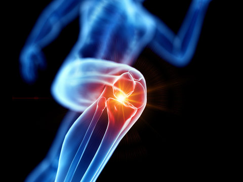
Infrapatellar Fat Pad Syndrome
Case study reflection blog
The purpose of this case study is to assist our graduates to fully reflect on a client that they have seen throughout the week. This will also form part of information that we can distribute to clients where they can read up on real life cases where we have been able to help clients and allow them to be pain free! Clients are refered to as Mr.X/Ms.X to keep their privacy.
Section 1: About your client and how you diagnosed the condition (think about how they presented, what subjective and objective information did you gather to help you diagnose?)
Ms. X presented to us with localised right knee pain but specifically worse over the anterior and medial aspect of the patella. The pain was described to be more of an ache rather than a sharp shooting pain which ruled out any neurological contribution, however, did report occasional clicking and catching. She further explained the pain wouldn’t be too bad in the morning but tends to worsen by the end of the day. Ms. X plays soccer outside of school for an elite team which involves x2 training session, x1 fitness session and game day. Ms. X also participates in basketball training and games at school as well as cross country. Activities that aggravate the right knee is passing during soccer, landing on it during basketball and after long periods of running. Due to the pain, the school basketball coach advised Ms. X to sit out of the game however no other activities have since been modified.
Upon examination, the sweep test was classified as grade 1 as the right knee appeared to be more swollen when compared to the left. Ms. X experienced pain during the McMurrays and Thessaly’s test. No pain was experienced during the Valgus and Varus Stress Test at 30o of knee flexion. Patella glides were also performed where gliding the patella posteriorly and medially elicited pain. During palpation, areas that triggered onset of pain included medial joint line and posterior aspect of the patella both on left and right side. Interestingly, a normal depth squat nor jump squats elicited pain, however, moving down into a deeper squat aggravated the knee.
Section 2: Your diagnosis and about the condition (what is your possible diagnosis?)
Possible diagnosis:
Infrapatellar Fat Pad Syndrome
Pathophysiology background:
The infrapatellar fat pad is located on the anterior aspect of the knee just behind the patella tendon. The specific function of this structure remains uncertain however, it’s believed to act as a reservoir for cells to repair the knee post-injury as well as shock absorption. The anatomical location of the infrapatellar fat pads causes it to be exposed to high mechanical loads, especially during knee extension. This structure has a rich blood and nerve supply causing increased sensitivity to pain in various knee conditions. Infrapatellar fat pad syndrome occurs when the fat pad becomes impinged between the patella and femoral condyle. This can lead to overuse or repeated microtrauma resulting in pain, swelling and inflammation.
Once swollen, the healing process becomes altered. If the fat pad is not given the opportunity to recover, it can become chronically inflamed which if not properly managed, fibrotic changes occur within the structure. Hence, repeated episodes of impingement drive a cycle of pain and swelling.
Section 3: Differential Diagnosis (what is another condition to consider and why?)
A meniscus injury (specifically medial meniscus), ligamentous tear, patellar tendonitis or patellofemoral pain could be a differential diagnosis for infrapatellar fat pad syndrome.
A medial meniscus injury could still possibly exist due to the positive result in both McMurrays, Thessaly’s and subjective report of occasional clicking and catching. However, this remains uncertain and requires further investigation next session. Ms. X’s right knee seemed to be quite inflamed, swollen and aggravated during the session and everything seemed to elicit pain. Once Ms. X allows some de-loading off that right knee and modifies sporting activities, it would be good to re-assess these tests to rule in or out a medial meniscus injury. Although regardless of the result, management and treatment would remain similar.
A MCL tear is unlikely, there was pain upon palpation of medial joint line however, when stressing MCL during Valgus Stress Test no pain was elicited and the ligament had a tight end feel with no gap in joint line.
Patellar tendonitis could be possible due to prevalence in basketball. However, pain is usually specific to the location of the tendon, rather than pain on each side of the tendon where fat pads are located and where Ms.X is reporting pain.
Patellofemoral pain causes more generalised pain on anterior aspect of the knee and often have no specific tender spots as the pain usually stems from behind the knee cap. Given Ms. X had specific pain and tenderness on fat pad location, patellofemoral pain remains less likely.
Section 4: Treatment (what did you do and why?)
My first session with Ms. X involved some manual therapy over the right quadricep with the aim to loosen the muscle and limit compression being forced onto the knee. To complement this, taping the knee to bias the patella to sit higher up was done to reduce the amount of compression and force being placed onto the infrapatellar fat pads to help reduce pain. Additionally, education was provided on the potential cause of this injury and how to manage it now but also prevent this from reoccurring in the future. Given that Ms. X is very active and participates in many sports, I re-assured her this injury can heal, it would just require some time to allow the inflammation and swelling to go down by modifying sporting activities and be able to listen to her body when it requires some rest. Ice, rest, and avoidance of aggravating activities was recommended for the time being. Lastly, prescription of strength-based exercises targeted at muscles surrounding the knee in the effort to help de-load the knee and allow other muscles to help manage the load evenly through the lower limb. The exercises included 2 sets of 40s wall sit x3, double leg glute bridge x8 and banded crab walks with clockwork x8. Quadricep and glute stretches for 30sx3 were also prescribed to prevent muscular tightness and improve infrapatellar fat pad restriction symptoms.
Section 5: Plan (where to go from here? How many sessions might they need? What’s the goal?)
Future sessions will be focused on progressing Ms. X through increasing set range and difficulty of strength-based exercises. I would also like to incorporate additional exercises focused on different lower limb muscle groups to achieve conditioning of the entire lower limb.
For the meantime, Ms. X is advised to cut back in some soccer and basketball training sessions unless able to participate in light passing or jogging activities. However, I would encourage full participation in fitness class the soccer club provides. In the future, I aim to get Ms. X back to participating in usual training sessions, but this will be done slowly overtime. Also, taping the knee is a strong recommendation I would still like to incorporate during the next couple of treatment sessions to help de-load the knee and reduce onset on pain.
I aim to review in one week, when the knee is less aggravated to allow me to distinguish more confidently between infrapatellar fat pad or medial meniscus injury. Given strength training takes ~8 weeks I would like to see Ms. X for a further 7 sessions. If she is coping well and starting to see improvements, we can potentially shorten this as she does participate in fitness sessions at soccer and is very active at school.

