
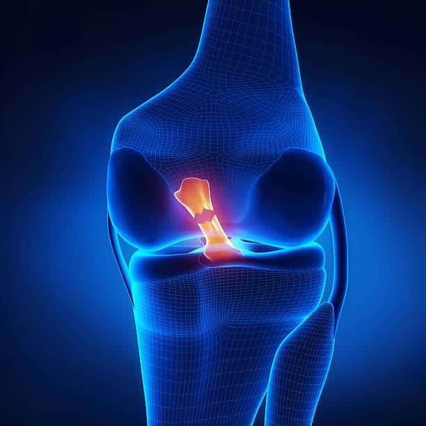
Medial meniscal tear
The purpose of this case study is to assist our graduates to fully reflect on a client that they have seen throughout the week. This will also form part of information that we can distribute to clients where they can read up on real life cases where we have been able to help clients and allow them to be pain free! Clients are refered to as Mr.X/Ms.X to keep their privacy.
TITLE OF BLOG: Medial meniscal tear
Section 1: About your client and how you diagnosed the condition (think about how they presented, what subjective and objective information did you gather to help you diagnose?)
Mrs. X presented to us with right knee pain following a netball match 10 days ago. She reported that she felt the pain after she twisted during a change of direction movement, and her knee has not been feeling 100% since. She is currently able to weight bear and walk, however the pain is aggravated when walking downstairs and sidestepping. She has not been taking any pain relief and she avoids/modifies aggravating factors to reduce her pain levels. Mrs. X describes the pain as being vague around and deep in the knee cap (patella), with an ache present. Mrs. X has a history of bilateral plantar fasciitis which she believes has been improving over the years. Mrs. X is a part-time school teacher and also has children of her own she looks after.
Upon examination, Mrs. X was tender upon palpation on both sides of the joint line of the patella, however more notably on the medial aspect. Anterior and posterior draw tests were conducted to assess the ACL and PCL integrity, with NAD L=R. A valgus and varus stress test was also performed to assess the integrity of the MCL and LCL, with findings also suggesting NAD L=R. The McMurray’s test was conducted and a positive finding on ER of the tibia with pain reproduced on the medial aspect of the knee joint. The Thessaly’s test was also performed with a positive finding on the medial aspect of the knee joint.
With the subjective and objective information gathered from Mrs. X, a medial meniscal tear was diagnosed.
Section 2: Your diagnosis and about the condition (what is your possible diagnosis?)
Possible diagnosis: Medial meniscal tear
Pathophysiology background:
The meniscus acts to increase stability for the femorotibial joint, as well as absorbing shock and distributing axial load from the other extremities.
A meniscal tear often occurs with flexion, compression, and femoral rotation, in which generates shear stresses that exceed the capabilities of the menisci. For example, a sudden change of direction in sports such as tennis and netball, or a direct impact to the knee causing twisting or rotation can stress the meniscus potentially causing injury.
In some cases, a meniscal tear can co-exist with ligament injuries of the MCL and ACL due to their attachments, and therefore a thorough assessment of the joint needs to be completed.
Section 3: Differential Diagnosis (what is another condition to consider and why?)
There are several differential diagnoses that are common in individuals with medial knee pain.
Collateral/cruciate ligament injury
A clinical presentation of an MCL injury is similar to that of a meniscal injury. Upon palpation, it is not uncommon for a patient with an MCL injury to report joint line tenderness, as well as a positive McMurray’s test. A positive valgus stress test could indicate an MCL injury and potentially rule out a meniscal injury. Additionally, the location of pain can vary between the two conditions, with an MCL injury usually causing symptoms above the joint line, and medial meniscal injuries displaying symptoms around and behind the knee. The mechanism of injury can also vary, with an MCL injury usually seen with a valgus force applied to a partially flexed knee.
PFJ related pain
The clinical presentation of PFJ related pain is also similar to meniscal injury. PFJ pain is often associated with a gradual insidious onset of pain in and around the patella, which is commonly aggravated by repetitive loading such as climbing up stairs, walking, and squatting. The patella grind test could be used to rule in potential PFJ related disorders.
Section 4: Treatment (what did you do and why?)
After assessing Mrs. X, we educated her on activity modification and movements to avoid in order to reduce her pain levels initially. We advised Mrs. X to not play netball for at least 1-2 weeks initially, as the sport involves twisting movements which could aggravate her symptoms and potentially cause further damage. We mentioned that movement within her pain threshold is encouraged and recommended to maintain her strength and mobility.
We then gave Mrs. X 4 exercises to complete for her home exercise program. The first exercise was straight leg raises to strengthen and build on her control of the quadriceps without excessively loading the knee. A slight ER of the ankle was also recommended to further bias the medial aspect of the muscle group. The next exercise involved a more functional sit-to-stand to again strengthen the quadriceps, as well as the hamstrings, gluteals, hip flexors and core muscles. The third exercise was a double leg glute bridge, with the aim to strengthen the gluteal muscles during hip extension. A theraband above the knees was also used to act as a reminder to keep the knees apart as well as engaging Mrs. X’s gluteals further. The final exercise was an isometric wall sit with 20-30 degrees knee flexion to again strengthen and condition the quadriceps.
Section 5: Plan (where to go from here? How many sessions might they need? What’s the goal?)
Future sessions will involve progressing Mrs. X’s exercises to target her neuromuscular control and global lower limb strength, with the aim to include plyometric and sport specific training and exercises. I can monitor Mrs. X’s progression by her symptoms as well as muscle strength through MMT and functional assessments such as a STS.
A study conducted by Lange et al. (2009) noted that lower limb strengthening exercises with a focus on quadriceps improves cartilage nutrition and can lead to a reduction in cartilage degeneration.
Soft tissue work through her major lower limb muscles could be beneficial for short term relief from muscle soreness as intensity and load increases during her rehab. It is important to take into consideration the increase in volume and load in order to reduce the likelihood of developing a tendinopathy.
Initially, it would be beneficial to check-in with Mrs. X once a week as the injury is in the acute phase and needs to be monitored.
With the suspected minor meniscal tear, the aim is for Mrs. X to return to netball in 4-6 weeks pain free and with confidence in the strength of her knee. If Mrs. X is still experiencing symptoms past the 6–8-week timeline, further imaging such as a CT scan or MRI may be warranted for further investigations.
References
Lange, A. K., Vanwanseele, B., Foroughi, N., Baker, M. K., Shnier, R., Smith, R. M., & Singh, M. A. F. (2009). Resistive Exercise for Arthritic Cartilage Health (REACH): A randomized double-blind, sham-exercise controlled trial. BMC Geriatrics, 9(1). https://doi.org/10.1186/1471-2318-9-1