Medial Tibial Stress Syndrome (Shin Splints)
What are Shin Splints?
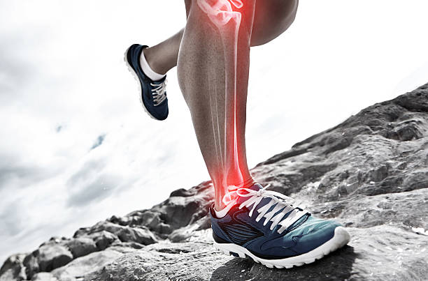
Shin splints are a common overuse injury to the lower limb. Commonly occurring in athletes and more specifically runners and military personnel. Shin splints cause pain over the anterior tibia and are considered an early-stage stress fracture. The incidence of shin splints is 13.6-20% in runners and 35% in military personal (Charles & Oh, 2019)
What causes this condition?
Shin splints is caused via an overuse mechanism. Patients who experience a high load and high impact through the lower extremities are typically most at risk of developing this condition. This high impact load through the distal tibia cause microtrauma of the tibia’s cortical bone and lead to inflammation of the periosteum. However, it is not yet clear in the research if the periostitis is caused by the microtrauma or if the microtrauma is caused by the periostitis (Franklyn & Oakes, 2015). Pain, in this case, is secondary to inflammation which results in increased traction of the Tibialis Posterior and soleus muscles, which both insert onto the proximal tibia.
Common symptoms / signs
Subjective:
Pain is present along the distal 2/3 of the medial tibial border during exercise and eases with relative rest. Inflammation along the medial border of the tibia is also common. Pain is at its most intense post-exercise bout (Winters et al., 2018)
Objective:
Pain on palpation of the posteromedial tibial border>5cm
Aggravators:
– Exercise
Eases:
– Relative rest
– Pain medications
– Ice Therapy.
Common Differential Diagnosis:
– Tibial Stress Fractures (common is the pain on palpation is less than 5cm)
– Chronic exertional compartment syndrome (Differentiating signs and symptoms: Cramping, burning in the posterior compartment, and/or neurological signs in the foot)
Risk factors
– Females
– PMHx of MTSS
– High BMI
– Navicular drops
– Reduced hip external rotation ROM
– Foot pronation
– Running on hard or uneven surfaces
– Triceps surae weakness of imbalance – causing additional GRF through the tibia
(Mattock et al., 2018)
How is it treated?
Treatment options within the current literature are scarce and have been completed with poor methodologies, therefore as clinicians, there is little evidence to guide our practice (Menéndez et al., 2020). However, exercise-based physiotherapy with the aim to relieve pain and allow patients to return to running without discomfort is the goal. Decreasing load off the passive bony structure of the tibia via relative rest from sports or running and adapting ADLs to decrease the load on the affected area, allowing pain to ease. Low-load cardio-based exercise such as hydrotherapy or cross trainers are important in the early stages to lessen the effects of a more sedentary lifestyle. Additionally, Knee and Hip driven exercises should be introduced accordingly, to strengthen the lower limb kinetic chain for optimal movement kinematics. Depending on the biomechanical deficits of the client these additional knees and hip exercises can be introduced earlier and/or running retraining may be required later in the rehab.
Ice therapy to reduce inflammation should be used in the acute stage of the pathology. However, CSI is contraindicated for analgesia due to healthy tissue also being affected in the procedure.
Sub-acute phase:
The main aim during this phase is to slowly reintroduce load into the client’s routine
Introducing lower limb stretching and strengthening exercises such as calf raises, banded dorsiflexion and eversion/inversion exercise is critical to beginning the strengthening process. Dosages and frequency should be tailored based on RPE and the client’s pain during exercise. However, we aim to undertake a muscular endurance protocol, as that closely resembles the athletes running profile, so higher repetitions are indicated.
Progress overload will take a similar approach, aiming to increase weekly to fortnightly without causing a reaction that aggregates the client. Progressions should aim to increase the transferability to running or the specific athletes’ sport as the program continues. Hence plyometric training should be included in the later stage of rehab before RTP.
This process of muscular and tendon strengthening can take anywhere between 6-8 weeks to see adaptions. These muscular adaptations will reduce the load of the tibia, decreasing pain innervated by the microdamage and secondary periostitis, relieving pain and ensuring appropriate bone healing can be completed.
Secondary lines of treatment such as Shockwave therapy, Dry needling and massage have not shown to have a minimal clinically important difference in the rehab of MTSS (Menéndez et al., 2020), additionally, these studies have had poor methodological quality. Despite this, they have also shown that they do not inhibit rehabilitation programs and therefore may be used as an adjunct If the client is agreeable. Foot orthoses to correct ankle inversion/eversion has been demonstrated to improve running kinematics (Naderi et al., 2019)
Reference:
Charles, J., & Oh, R. (2019). Medial Tibial Stress Syndrome Pathophysiology Treatment / Management. 1–5.
Franklyn, M., & Oakes, B. (2015). Aetiology and mechanisms of injury in medial tibial stress syndrome: Current and future developments. World Journal of Orthopedics, 6(8), 577–589. https://doi.org/10.5312/wjo.v6.i8.577
Mattock, J., Steele, J. R., & Mickle, K. J. (2018). A protocol to prospectively assess risk factors for medial tibial stress syndrome in distance runners. BMC Sports Science, Medicine and Rehabilitation, 10(1), 1–10. https://doi.org/10.1186/s13102-018-0109-1
Menéndez, C., Batalla, L., Prieto, A., Rodríguez, M. Á., Crespo, I., & Olmedillas, H. (2020). Medial tibial stress syndrome in novice and recreational runners: A systematic review. International Journal of Environmental Research and Public Health, 17(20), 1–13. https://doi.org/10.3390/ijerph17207457
Naderi, A., Degens, H., & Sakinepoor, A. (2019). Arch-support foot-orthoses normalize dynamic in-shoe foot pressure distribution in medial tibial stress syndrome. In European Journal of Sport Science (Vol. 19, Issue 2, pp. 247–257). https://doi.org/10.1080/17461391.2018.1503337
Winters, M., Bakker, E. W. P., Moen, M. H., Barten, C. C., Teeuwen, R., & Weir, A. (2018). Medial tibial stress syndrome can be diagnosed reliably using history and physical examination. British Journal of Sports Medicine, 52(19), 1267–1272. https://doi.org/10.1136/bjsports-2016-097037
